
15 Aug Angiography method
Angiography method
Angiography is an excellent diagnostic method for examining the coronary arteries of the heart as well as the arteries of other parts of the body and determining the degree of vascular obstruction and vessel problems. To perform angiography, the patient is admitted to a hospital equipped with an angiography machine and after the tests performed, he is transferred to a special room to perform angiography. After sterilizing the groin area (or any area to be performed from there), a suitable cover is placed on the patient and the patient lies motionless on a bed equipped with a fluoroscopy (X-ray imaging) machine. In angiography, usually the femoral artery is pierced in the groin area after local anesthesia and a very thin tube (sheath) is placed in the artery and then very thin tubes (very thin catheters) are inserted through the angiography sheet.
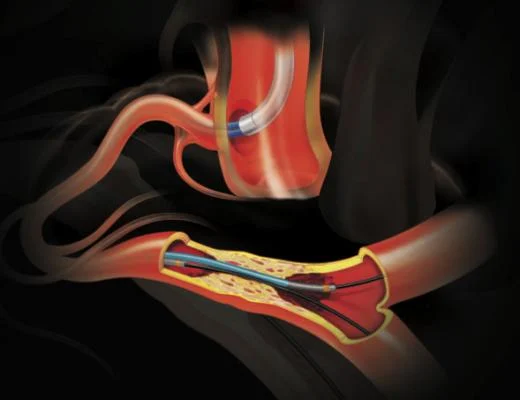
During the angiography, diagnostic catheters (50 cm long) are inserted into the artery from inside the sheet and guided by fluoroscopy into the coronary arteries (the main arteries of the heart) or any other part that is to be subjected to angiography, in picture 1 1- The stages of hagiography are visible. After the catheter is placed at the beginning of the vein, the contrast material (special liquid) enters the vein through the catheter and simultaneous imaging is done using X-ray. As in picture number 1-2, the vein and its disorders are displayed on the monitor (screen). ) is displayed and recorded.
Contrast injection and imaging were repeated as many times as required, and after the completion of the angiography, the catheter as well as the tubular instrument (sheet) was removed from the patient’s leg, and one of the angiography personnel, with his hand, for several minutes on the artery from which the sheet It is removed, pressure is applied so that bleeding does not occur.
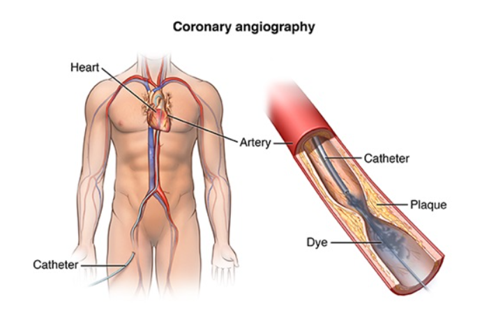
Figure 1-1, Angiography steps
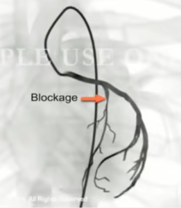
Picture 1-2, showing the location of the vein blockage
Complications of angiography
Angiography (cardiac angiography or angiography of vessels of other organs) usually does not have any complications, in some cases, minor side effects such as nausea and vomiting may occur, which are quickly resolved by prescribing medication. Angiography of the heart or any other part of the vessels is usually done through the groin veins (femoral artery), although it can also be done through the hand veins.
The operation of angiography of the heart or other vessels of the body is low risk with the available facilities, and side effects such as mild pain in the angiography area usually resolve quickly. A special skin lesion does not remain at the angiography site.
After angiography
It should be noted that angiography is only a diagnostic method and does not lead to the opening of blood vessels. Based on the vascular lesions observed in angiography, one of the three options of balloon angioplasty, surgery (CABG) or drug treatment may be chosen for the patient. If the conditions are suitable, balloon angioplasty may be performed immediately after angiography (i.e. in the same session), but sometimes it is postponed to the next session due to special reasons. During angiography, at the same time that the patient is lying on the bed, the angiography imaging camera, by rotating certain degrees around the patient, takes images from different angles, so any lesion in the vessels can be detected using angiography.
No Comments