logo
Copyright Pouyan Tajhiz 2016 Designed by Pulse Design
 

URF-3AP+MLA-4

products-feature-3AP-mla4
products-feature-ml4-history
products-feature-ml4-lesion-2bp
products-feature-ml4-lesion-c
products-feature-ml4-lesion-p
products-feature-ml4-lesion-quad
products-feature-ml4-lesion-stg
products-feature-ml4-probe-test
products-feature-ml4-stim-motor
products-feature-ml4-stim-sens

One of the challenges is treating back pain is the many possible sources of the pain. Pain in the low back or buttock may arise from muscle, intervertebral disc, facet joints, sacroiliac joints, o ligaments. In 1911, Goldthwait recognized the facet joint as a potential source of pain. Rees in 1971 first reported on surgical facet joint denervation by the passage of a tentomy blade to the inter transverse ligament to section branches of the posterior primary ramus. In 1974, Shealy introduced percutaneous radiofrequency facet joint denervation but his results were compromised by the use of an incorrect target location, distant from the now accepted target of the medical branch of the posterior primary ramus as it courses I the groove formed by the junction of the transverse process and superior articular process.

There is no radiographic or physical examination that can unequivocally identify a facet joint as the source of pain. However, a specific diagnostic test that has evolved is the assessment of the location and duration of pain relief following an anesthetic block of the medical branch nerve. A result that is predictive of successful RF ablation of the medical branch is about 70 to 80% pain relief lasting the expected duration of the anesthetic agent.

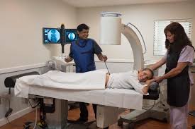
The advences in digital fluoroscopy and mobile C-arm technology have greatly enhanced the ability to perform these RF procedures with precision and safety. The procedures can be performed on an outpatient basis under local anesthesia and when necessary, under conscious sedation. In addition, the use of ultrasonic imaging as a guidance technique is being explored, but at present a limitation of this modality is the depth of structures that must be visualized the lumbar area.

The Diros OWL® URF-3AP Lesion Generator with Multilesion Adapter (MLA-4) independently controls up to four RF Probes at the same time. With the ability of catering to the user’s requirements the following features are available:

  • Impedance monitoring during all modes and for up to 4 channels simultaneously.
  • Stimulation: sensory and motor stimulation available in constant voltage or current modes.
  • RF Lesion modes: Continuous or pulsed monopolar, bipolar, dual bipolar, or patent pending QuadrapolarTM.
  • Automatically controlled RF output with simultaneous or staggered starts, or manual RF output control.
  • Integral RF Probe and Cable tester allows user to check cable and probe functionality, including electrical continuity, power delivery, and temperature accuracy.
  • History Recorder with output to a USB flash drive allows the user to save the last 127 procedures performed.
  • Customized user presets allow for up to five users to save and store display and procedure parameters as their default settings with optional password protection.
  • Language selection, over 17 languages to choose from such as: English, French, German, Greek, Hindi, Hungarian, Italian, Japanese, Korea, Portuguese, Russian, Spanish, Thai, Turkish, Chinese (Simplified and Traditional), Dutch.
  • Custom carrying case available for stylish and secure transportation


images

Facilities and Equipment

A facility for interventionism spine procedures should include:

A sterile procedure room equipped with a high quality digital C-arm fluoroscopy unit and viewing screen.

X-ray protective lead aprons and thyroid shields.

A radiolucent procedure table.

One pillow to place under the head for comfort while in the prone position, and another under the lower abdomen to flatten the lumbar lordosis and tilt the pelvis posteriorly.

Monitoring equipment for blood pressure,pulse oximetry, and ECG.

Resuscitation equipment.